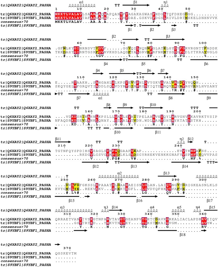
Figure 4.

Alignment between MhHM [UniProtKB: Q6XAY2] and MhMP sequences [UniProtKB: S9YBF1]. The prediction of the secondary structure performed with PSS PRED is shown in the top and the bottom of each sequence; the arrows show the β-sheets and the spiral α-helix structure. The red boxes show amino acid identity and the yellow boxes similarity [40, 41].
