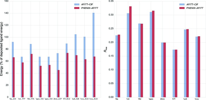
Figure 5.
Comparison of eight randomly selected PDB structures. The left panel shows energies obtained with AFITT–CIF refined and PHENIX–AFITT refined ligand restraints as a percentage of the deposited ligand energy. Labels provide the PDB code followed by the three-letter code for the ligand. Some PDB structures have more than one instance of a ligand. The right panel shows the R free obtained after refinement with Engh and Huber or AFITT geometry restraints on the ligands.

