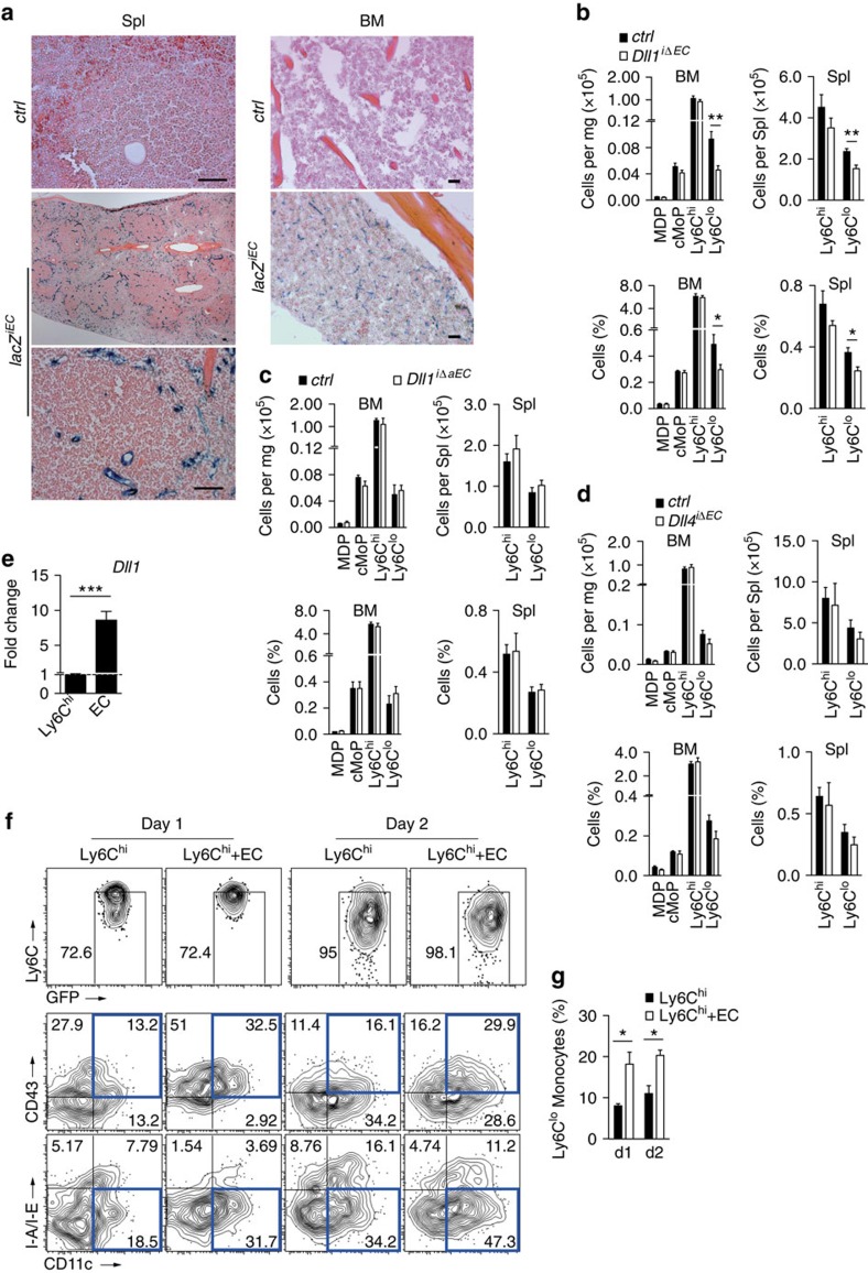
Figure 8. Endothelial deletion of Dll1 impairs Ly6Clo monocyte development in vivo.
(a) Inducible and endothelial-specific Cre-reporter mice (lacZiEC) showing specific β-galactosidase staining in splenic MZ (left) and BM (right) of the femur diaphysis after tamoxifen treatment; scale bars, 50 μm. (b) Absolute and relative numbers of myeloid cell populations by flow cytometry after pan-endothelial deletion of Dll1 compared with littermate controls. Data are pooled from four experiments (n=8). (c) Absolute and relative numbers of myeloid cell populations by flow cytometry after arterial-specific, endothelial deletion of Dll1 compared with littermate controls. Data are pooled from two experiments (n=5/6). (d) Absolute and relative numbers of myeloid cell populations by flow cytometry after endothelial-specific deletion of Dll4 compared with littermate controls. Data are pooled from two experiments (n=5). (e) Quantitative reverse transcription–PCR analysis of Dll1 expression in sorted splenic monocytes (CD11b+GFP+Ly6ChiCD144−) and ECs (CD11b−GFP−CD144+). Data are pooled from three-independent experiments (n=6). (f,g) Co-culture of splenic CD144+ ECs with Ly6Chi monocytes. Representative flow cytometry plot from GFP+CD11b+ gate (f) and frequency of Ly6Clo monocyte-like cells (g) are shown (n=3). *P<0.05, **P<0.01, ***P<0.001; Student's t-test (b–e) or one-way ANOVA with Bonferroni's multiple comparison test (g). (b–e,g) Error bars represent s.e.m.

