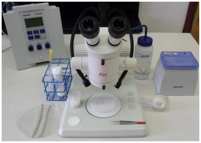
Figure 1. A prepared work station ready for lining up embryos for microinjection.
Upon the desk from the left then proceeding clockwise: needles within their container, a laying chamber tube and a 15 ml tube of Halocarbon Oil with a glass Pasteur pipette ready for covering dessicated embryos, the Eppendorf Femtojet, a water bottle with a fine jet spray nozzle, Eppendorf microloaders (within their tip-rack) and double-sided adhesive tape. Upon the microscope stage (from left to right) are a small circle of paper for the laying chamber, a recovery beaker, a Whatman paper circle upon a petri dish lid showing a line of embryos ready for transfer, a pair of tweezers and three prepared coverslips.

