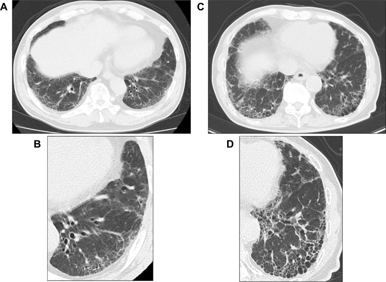
Figure 2.
High-resolution computed tomography (HRCT) images demonstrating usual interstitial pneumonia (UIP) pattern and possible UIP pattern. (A and B) UIP pattern, with extensive honeycombing: conventional and HRCT images show basal-predominant and peripheral-predominant reticular abnormality with multiple layers of honeycombing. (C and D) Possible UP pattern: conventional and HRCT images show peripheral-predominant and basal-predominant reticular abnormality with a moderate amount of ground glass abnormality, but without honeycombing.

