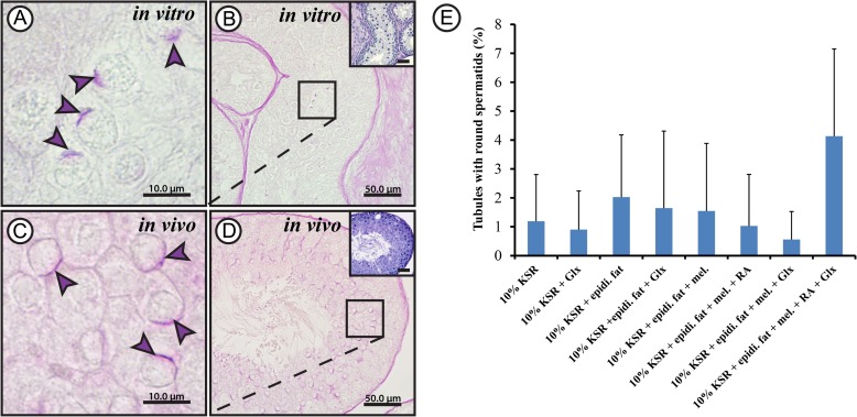
Figure 3.
Morphological evaluation results for round spermatids obtained from rat testicular tissue culture. Periodic acid Schiff's staining (PAS) of Bouin's solution-fixed paraffin-embedded testicular tissue cultured for 52 days in minimum essential medium alpha (MEMα) + 10% (v/v) knock-out serum replacement (KSR) (A and B) and 60 days postpartum adult rat testis control (C and D) showing round spermatids. Higher magnifications of B and D are shown in A and C, respectively. Hematoxylin was used for counter-staining in the same samples to show overall organization in the tissue (small inserts; B and D). The violet arrow heads show acrosomal caps. The scale bars are 10 µm (A and C) and 50 µm (B and D). Percentage of tubules containing spermatids exhibiting acrosomal caps (E) compared between the different culture conditions used; MEMα, minimal essential medium α;KSR, knock-out serum replacement; Glx, Glutamax; epidi. fat, epididymal fat; mel, melatonin; RA, retinoic acid. Values are mean ± SD (n = 6–12). For statistical analysis, one-way ANOVA test was applied to compare between the percentages of tubules from the different culture conditions.

