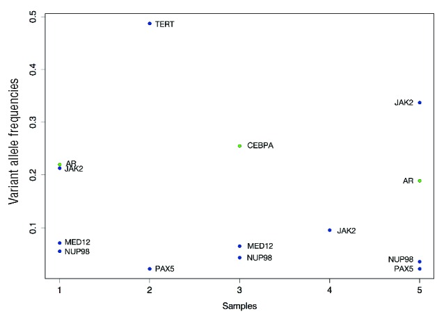
Figure 1.

Genes mutated in pediatric ET patients. The figure illustrates the genes mutated for each of our samples with the corresponding variant allele frequencies. Insertions and deletions are colored green and single nucleotide variants are colored blue.
