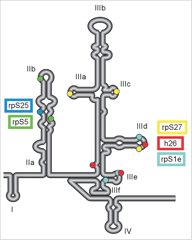
Figure 1.

Multiple contact points between HCV IRES and 40S ribosomal subunit. Schematic diagram of the secondary structure of HCV 5′ UTR. Four distinct domains (I-IV) are labeled along with subdomains of IIa-b and IIIa-f. The IRES regions that were identified to interact with various components of 40S subunit by CryoEM analysis are indicated with color-coded dots: rpS5 (uS7; green), rpS25 (blue), rpS27 (yellow), rpS1e (cyan) and 18S rRNA helix 26 (red).40,54. Additional nucleotides 70-74 and 84-91 of HCV IRES are predicted to make contact with rpS5.39
