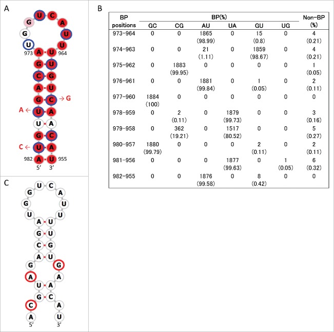
Figure 4.
Stem loop structure of SL5–3B(+) predicted at nucleotide positions 967–994 of M segment +RNA. (A) Secondary structures were predicted using the M nucleotide sequence of England/195/2009(H1N1) (accession number GQ166660) at temperature setting of 37°C. The background red, pink and white colors of each base represents nucleotide identities of > 98%, 90–98%, and< 90% among 1,884 sequences, respectively. Third codon positions are circled in blue. The red arrows and nucleotide sequences indicate the position and nature of synonymous mutations that were introduced to produce SL5–3B mutants. (B) The number (percentage) of sequences that were predicted to form base-paring (BP) at the stem positions of SL5–3B(+) among 1,884 sequences. (C) Secondary structure at positions 967–994 of SL5–3B M sequence. Bases with synonymous mutations are circled in red.

