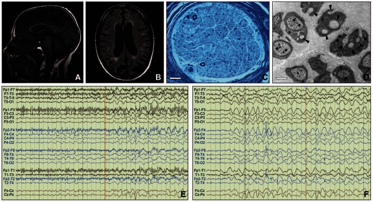
Figure 2.
Imaging results. (A) Brain MRI in Patient III-2 from Kindred 7, a 54-year-old male with significant cognitive impairment and diffuse slowing on EEG: sagittal T1 image showing global supra- and infra-tentorial brain atrophy (Philips, 1.5 T). (B) Transversal FLAIR image showing multiple white matter lesions and global atrophy (Philips, 1.5 T). (C) Morphological analysis of glutaraldehyde-fixed sural nerve biopsy tissue embedded in epoxy resin of Patient II-1 from Kindred 3: showing severely reduced numbers of myelinated nerve fibres. Semi-thin section, toluidine blue. Scale bar = 25 µm. (D) Remak bundles containing several bundles of collagen fibres encircled by Schwann cell processes (collagen pockets, indicated by arrowheads) indicative of unmyelinated axon loss. Electron microscopy of ultrathin section. Scale bar = 1 µm. (E) Twenty-four hours of continuous video-EEG monitoring in Patient II-2 from Kindred 1 showing irregular myoclonic movements associated with runs of higher voltage, irregular theta activity. (F) EEG in the same patient showing abnormal diffuse rhythmic delta frequency (1–4 Hz) background slowing especially in the bifrontal regions, correlated with a mild to moderate diffuse encephalopathy.

