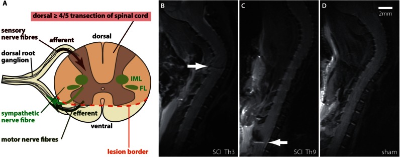
Figure 2.
Experimental SCI with differential lesion levels (Th3 versus Th9) of the sympathetic preganglionic neurons. (A) The CNS controls the immune system through the autonomous nervous system. SCI specifically disconnects sympathetic preganglionic neurons located in the thoracolumbar spinal cord, which control systemic immune function by postganglionic noradrenergic projections to secondary lymphoid tissue including the spleen. Inhibition of spinally generated SNA and reflex excitation through sympathetic preganglionic neurons by baroreceptors and other supraspinal inhibitory systems (bulbo-spinal tracts) is lost after complete SCI above mid-thoracic level (‘isolated sympathetic spinal cord’ by ‘sympathetic decentralization’). Episodes of excessive SNA correlate with injury severity and lesion level. Lesion level dependent injury of sympathetic preganglionic neurons localized to the inter-mediolateral columns (IML) and funciculus lateralis (FL) in the thoracic rodent spinal cord is assured by a dorsal > 4/5 transection SCI. (B–D) Severity, consistency and SCI lesion level were confirmed by MRI 1 day before infection. Animals with spared ventral tissue bridges were excluded. Representative T2 MRI images, illustrating a spinal cord lesion at the respective SCI level Th3 (B) and Th9 (C) (white arrows) compared with the uninjured spinal cord after sham surgery (D).

