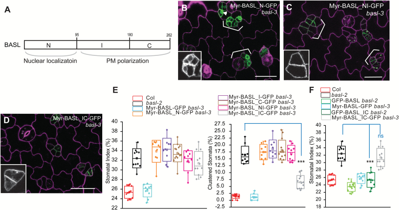
Fig. 5.
Localization and function of myristoylated BASL domains.
(A) Box diagram shows BASL subdomains, N (N-terminal), I (Internal), and C (C-terminal). BASL_N was found to direct nuclear localization and BASL_IC mediates polarization at the PM. (B–D)Confocal images show localization of fusion proteins in basl-3 (green), Myr-BASL_N-GFP (B), Myr-BASL_NI-GFP (C), and Myr-BASL_IC-GFP (D). Cells were outlined with PI staining (magenta). Representative protein localization is shown with the GFP channel only (insets). Brackets highlight stomatal clustering. Note the Myr-BASL_N-GFP protein in the cytoplasm and some in the nucleus (inset in B), likely due to the antagonistic localization mechanisms to the nucleus and to the cortical membrane. Scale bar = 25 µm. (E) Box plots show quantification of stomatal phenotypes in complementation. Myr-BASL_IC partially rescued basl, in particular in terms of clustered stomata (Student’s t test, ***P < 0.0001 compared to basl-2). (F) Box plots show the comparison of transgene complementation. GFP-BASL_IC, but not Myr-BASL_IC-GFP, most effectively rescued the basl null mutant. Mann–Whitney test, *** P < 0.001 compared to basl-2 and ‘ns’ indicates ‘not significant’.

