
Figure 1.
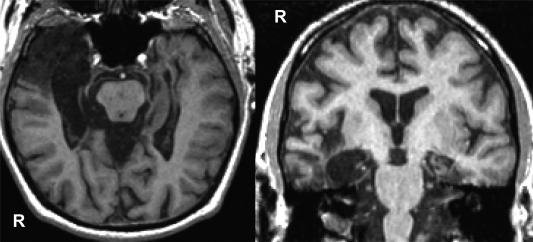
T1‐weighted images in axial (left) and coronal (right) views showing lesions to bilateral medial temporal lobe and right anterior temporal cortex in DA.
Official websites use .gov
A
.gov website belongs to an official
government organization in the United States.
Secure .gov websites use HTTPS
A lock (
) or https:// means you've safely
connected to the .gov website. Share sensitive
information only on official, secure websites.
T1‐weighted images in axial (left) and coronal (right) views showing lesions to bilateral medial temporal lobe and right anterior temporal cortex in DA.