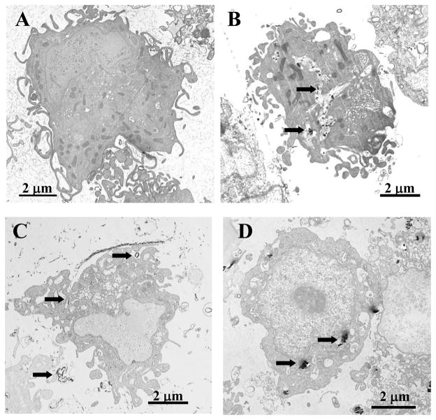
Figure 3.
Cellular uptake and ultra-structural effects of fibrous nanomaterials. Transmission electron micrographs of RAW264.7 macrophages after exposure to CNF (B), SWCNT (C) or asbestos (D). PBS-exposed control cells are shown in (A). RAW264.7 macrophages were incubated with 24 μg/cm2 of CNF, asbestos or SWCNT for 24h (37°C). Arrows indicate engulfment of the particles.

