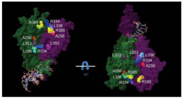
Figure 1.
General view of a NR2E3 homodimer bound to DNA. The crystal structure of the HNF4α/NR2A1 (isoform a) homodimer bound to DNA (PDB_4IQR) (Chandra, et al., 2013) served as a template to superpose the structure of the NR2E3 LBD dimer (PDB_4LOG) in an auto-repressed conformation (Tan, et al., 2013). One monomer is shown in dark green, the other in dark purple, both contacting the DNA double helix. ESCS-linked residues located close to the dimer interface are indicated in colors: A256 in red, R334 in blue, L336 in cyan, L353 in magenta and R385 in yellow. To facilitate visualization, two different views turned by 90°C are shown. For clarity, residues R309 and R311 that are not predicted to be in close proximity of the dimer interface are not indicated (see Supp. Figure S1).

