Figure 5.
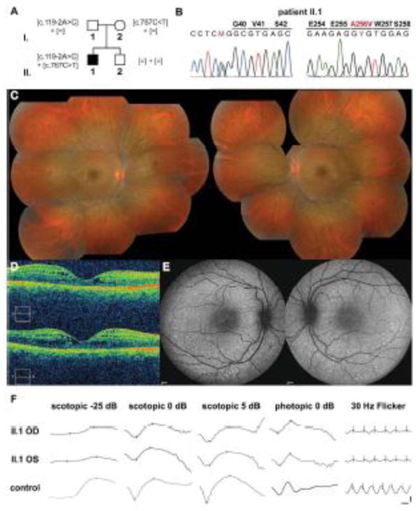
Compound heterozygous NR2E3 [c.119-2A>C];[p.A256V] patient. A) Pedigree of the Italian family with recessive inheritance of the [c.119-2A>C] variant from the unaffected father and the [c.767C>T] (p.A256V) variant from the unaffected mother. None of the variants was detected in the unaffected brother II.2. B) Electropherograms of the heterozygous substitutions c.119-2A>C in exon 2 and c.767C>T (p.A256V) in exon 6 present in patient II.1. Sequences were aligned with the reference genomic NR2E3 sequence NW_001838218.2. C) Composite fundus photograph of the 18-year-old proband II.1. Small white/yellowish dots and flecks along the vascular arcades and around the macula are observed (OD: left panel; OS: right panel). D) Optical coherence tomography (OCT) of the right (upper panel) and left eyes (lower panel) of the proband. Foveal schisis is visible in both eyes. E) Fundus autofluorescence examination revealed hyperautofluorescent pin-points along the vascular arcades in both eyes (OD: left panel; OS: right panel). F) ISCEV scotopic and photopic full-field ERGs revealed residual rod-specific responses (scotopic -25 dB), decreased scotopic maximal responses (scotopic 5 dB), delayed transient photopic responses with normal amplitudes (photopic 0 dB). The photopic 30-Hz flicker was delayed and of decreased amplitude. Horizontal bar: 20 msec; vertical bar: 50 μV.

