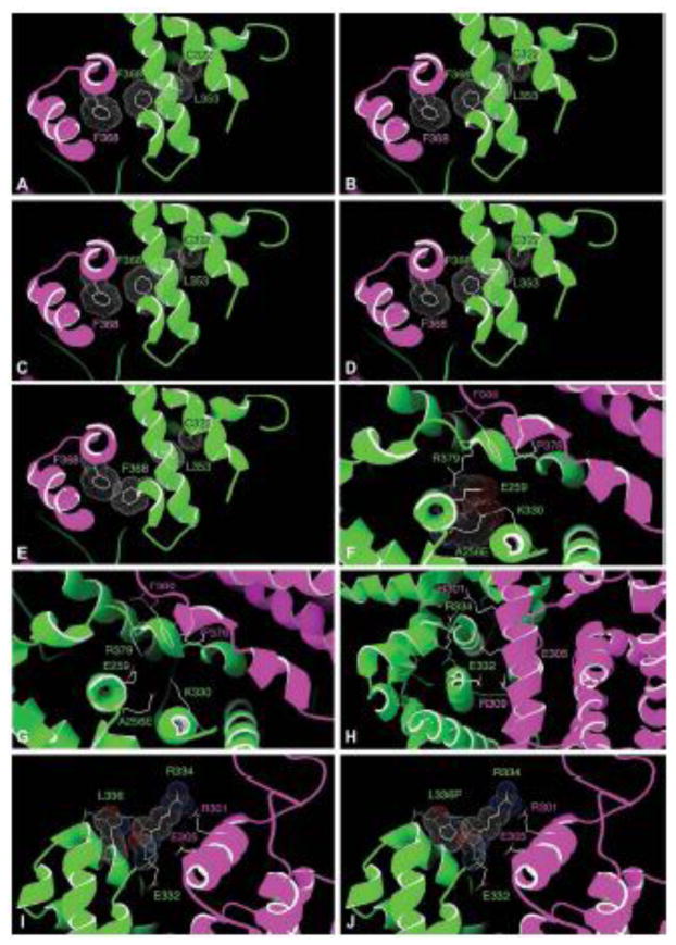
Figure 6.
Structural contexts of disease-causing NR2E3 LBD variants based on the model shown in Figure 1 (see discussion). Detailed views of the contexts of the A256 residue (A–B), the R334 residue (C), the R334 and L336 residues (D–E) and the L353 residue (F–J). One NR2E3 LBD monomer is shown in purple, the other in green. For clarity, only the two last helices of the green monomer are shown in E–H.

