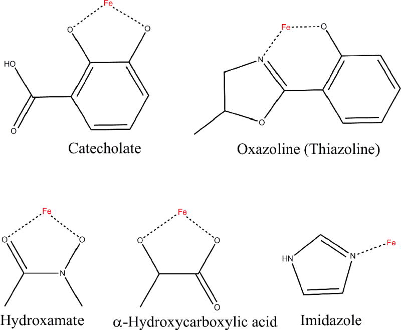
Fig. 5.

The functional groups of siderophores for chelating iron.
The protein images were made with VMD software support. VMD is developed with NIH support by the Theoretical and Computational Biophysics group at the Beckman Institute,University of Illinois at Urbana-Champaign http://www.ks.uiuc.edu/Overview/acknowledge.html.
The structure of ligands (1–62) were drawn by chembiodraw ultra 12.0
