Figure 1.
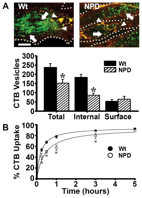
Binding and internalization of cholera toxin B (CTB) in NPD fibroblasts. A Fluorescence microscopy of wild type (Wt) and NPD fibroblasts incubated with CTB (green) for one hour at 37°C. Unbound CTB was washed off, cells were fixed, and surface CTB was immunostained (red+green=yellow; arrowheads) to distinguish it from internalized CTB (green; arrows). Dotted lines = cell borders, as observed by phase-contrast microscopy. Scale bar = 10 μm. Total, internal, and surface CTB (fluorescent objects over background) were quantified. B Kinetics of uptake of CTB in Wt vs. NPD fibroblasts, examined as described in A, where the percentage of internalized fluorescence from the total cell-associated fluorescence is shown. Lines are regression curves, where R2 was 0.99 for Wt and 0.94 for NPD. A–B Data are the mean ± SEM. *Comparison with Wt cells (p<0.05 by Student’s t-test).

