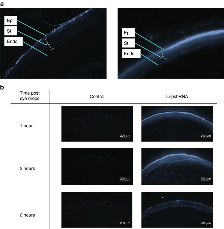
Figure 2.
Delivery of fluorescence-labeled pshRNA eye drops. (a) Left panel: bare siRNA with fluorescent label; right panel: fluorescence-labeled pshRNA enclosed in the lipid nanoparticle. The pshRNA was distributed only in the superficial layer of the corneal epithelium, while the Li-pshRNA was observed in almost all layers. (b) Delivery of fluorescence-labeled Li-pshRNA drops to mouse eyes. Samples were extracted at 1, 3, and 6 hours after instillation of the eye drops. Left panel: saline as control. Right panel: fluorescence-labeled Li-pshRNA. The Li-pshRNA was distributed to all layers as time advanced. Epi, epithelium; Endo, endothelium of cornea; St, stroma. Li-pshRNA, proline-modified short hairpin RNA wrapped with liposome.

