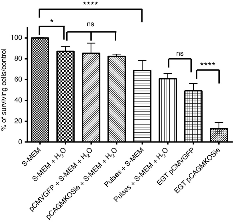
Figure 2.
Specific toxicity of each component of the electrotransfer protocol for a small and a large plasmid. Adipose tissue-derived mesenchymal stem cells were exposed to each component of the electrotransfer alone or in combination: pulsing buffer (S-MEM), improved pulsing buffer (S-MEM with 50% H2O), small (pCMV-GFP, 3.5 kbp, 50 µg) or large plasmid (pCAGMKOSiE, 11.4 kbp, 50 µg), eight electric pulses (1,500 V/cm, 100 µs, 1 Hz), complete EGT (S-MEM + 50% H2O + plasmid + pulses). For each condition, cells were counted with the flow cytometer 24 hours after treatment and survival expressed as the percentage of cells counted in the control (S-MEM). Data are representative of three to five independent experiments. (*P < 0.05 and ****P < 0.0001, one-way analysis of variance with Holm-Šídák multiple comparison test). CMV, cytomegalovirus; EGT, electrogene transfer; GFP, green fluorescent protein; ns, nonsignificant; S-MEM, minimum essential medium modified for suspension cultures.

