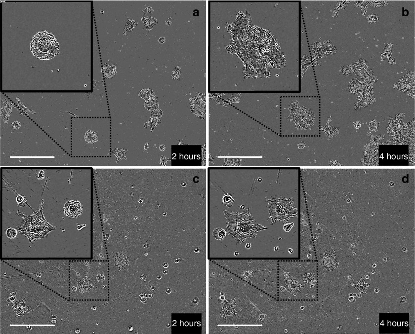
Figure 4.
Toxicity of small and large plasmid 2 and 4 hours after electrotransfer. Adipose tissue-derived mesenchymal stem cells were electrotransferred (eight pulses, 1,500 V/cm, 100 µs, 1 Hz) with 50 µg of either the small pCMV-GFP plasmid (3.5 kbp) (a,b) or the large pCAGMKOSiE plasmid (11.4 kbp) (c,d) and observed 2 hours (a,c) and 4 hours (b,d) after electrotransfer using a phase contrast objective. Within less than 2 hours, the cells were attaching and at 4 hours, they were well spread on the surface. For large plasmid, a significant amount of cells did not attach and some were blebbing. Bar = 400 µm. Pictures are representative of three experiments. CMV, cytomegalovirus; GFP, green fluorescent protein.

