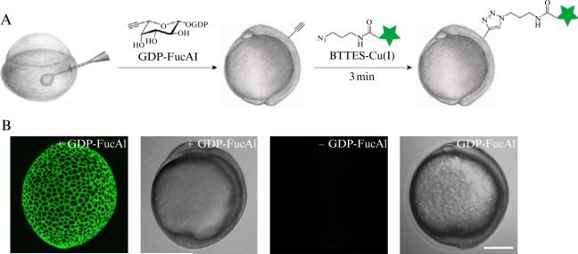
Figure 21.3.
A schematic description of metabolic labeling of cell surface fucosylated glycans in zebrafish embryos for fluorescence imaging. (A) workflow of metabolic labeling of fucosylated glycans followed by conjugation with fluorophores via CuAAC for imaging; (B) representative images of zebrafish embryos treated by this process: from left to right, fluorescence image of 10 hpf embryos treated with GDP-FucAl followed by a click reaction with Alexa Fluor 488-azide; the corresponding bright field image of 10 hpf zebrafish embryos treated with GDP-FucAl followed by a click reaction with Alexa Fluor 488-azide; fluorescence image of 10 hpf embryos treated without GDP-Fuc; the corresponding bright field image of 10 hpf embryos treated without GDP-Fuc followed by a click reaction with Alexa Fluor 488-azide.

