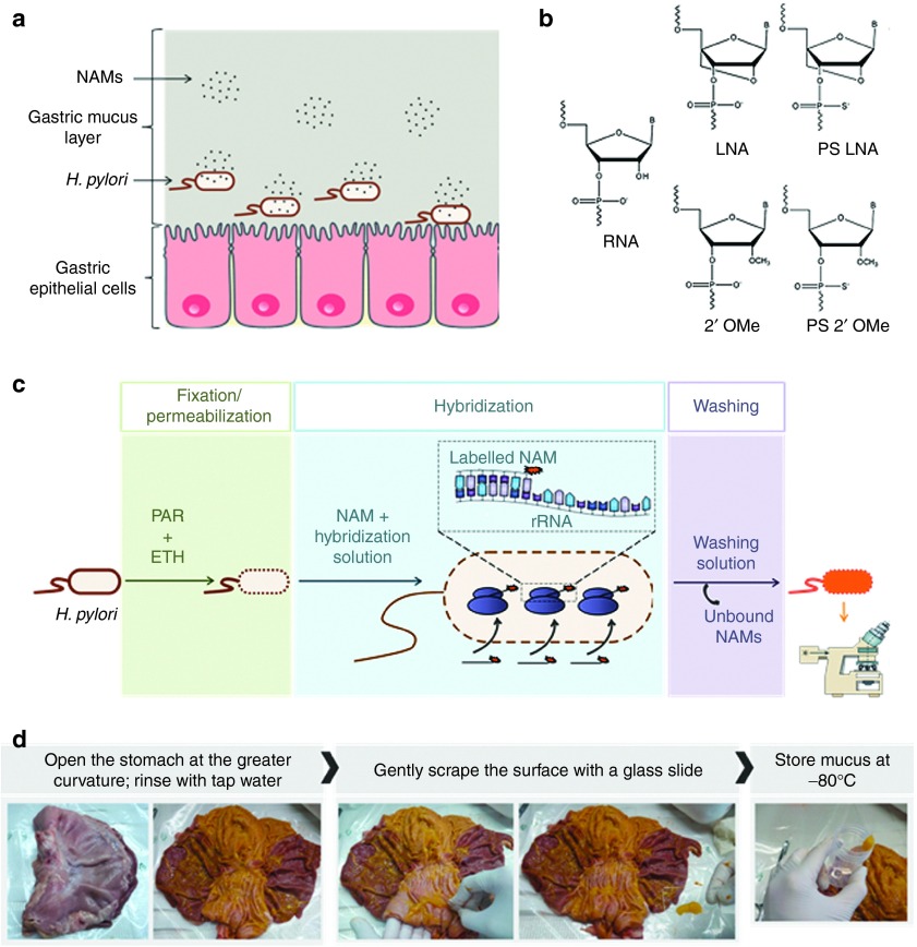
Figure 1.
Illustration of nucleic acid mimics (NAMs) hybridization to H. pylori and the different components of the implemented model. (a) Schematic representation of NAMs in gastric mucus, on their way to target H. pylori. (b) Monomers of the NAMs used in fluorescence in situ hybridization (FISH), compared to RNA (adapted from32). (c) Illustration of the standard FISH procedure. PAR and ETH being paraformaldehyde and ethanol, respectively (adapted from ref. 30). (d) Procedure followed for the collection of native mucus from the stomach of pigs, obtained from the slaughterhouse. ETH, ethanol; PAR, paraformaldehyde.

