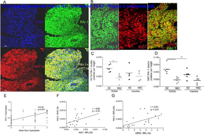
Figure 2.

Actively proliferating cells express PAI-1. Representative images of double immunofluorescence for PAI-1 (green, A, B) and cytokeratin 14 (Krt14) (red, A) or nuclear Ki67 (red, B) shows that PAI-1 co-localizes with cytokeratin 14 (yellow, A) and Ki67 positive cells (green cytoplasm, red nucleus, B). Quantification of cells that are KRT14-PAI-1 (C) and Ki67-PAI-1 (D) double positive in the basal (BZ) and non-basal (NBZ) zones of a subset of biopsies. Epithelial PAI-1 expression correlates with the severity of basal zone hyperplasia (E). PAI-1 mRNA expression correlates with Ki67 (F) and cytokeratin 5 (Krt5) (G) in esophageal biopsies from active, inactive, and control subjects.
