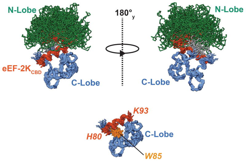
Figure 5. Solution structure of the Ca2+-CaM•eEF-2KCBD complex.
20 structures representing the final NMR ensemble have been overlaid using the C-lobe and eEF-2KCBD (see Table 1). The N-lobe (1–77) and C-lobe of CaM (82–148) have been colored green and blue respectively and the linker (78–81) is colored grey. eEF-2KCBD is colored dark orange. The N-lobe of CaM, the linker, 6 residues on the N-terminus and 7 residues on the C-terminus of eEF-2KCBD are hidden to allow better visualization of the C-lobe eEF-2KCBD interactions in the lower panel. The sidechains of the critical W85 are shown and colored light orange. The eEF-2KCBD residues are in italics and shown in orange. The Ca2+ ions occupying the N-lobe sites are not shown.

