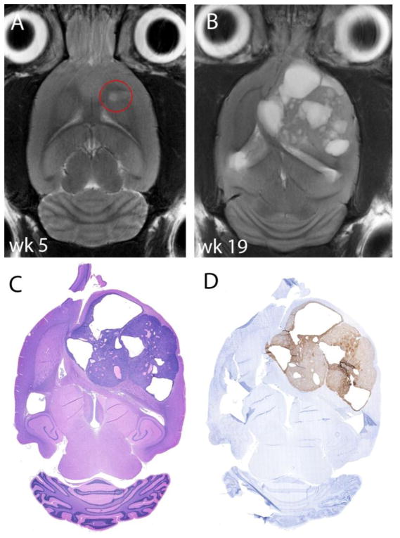
Figure 6.
MRI images and histology of a control animal who did not receive treatment. A) Tumor on T2w imaging 5 weeks after implantation. B) Tumor on MR imaging when the animal reached the study endpoint. The tumor shows a heterogeneous appearance with cystic cavities. C) The hematoxylin & eosin-stained section corresponds well with MR-imaging and cysts are present. D) The HER2-stained section demonstrates that the complete tumor is HER2-expressing.

