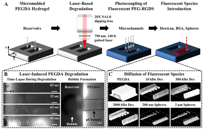
Figure 1. Fabrication of Microfluidic Channels in PEGDA Hydrogels.
(A) A 5% 3.4 kDa PEGDA hydrogel is photopolymerized against a PDMS master to create reservoirs. A 790 nm, 140 fs pulsed laser focused through a 20X(NA1.0) water immersion objective is raster scanned in desired 3D configurations to locally degrade the hydrogel. The hydrogel is fluorescently labeled via photocoupling of a monoacrylate fluorescent PEG-RGDS to visualize degraded volumes. The reservoirs are filled with a fluorescent species (dextran (Dex), bovine serum albumin (BSA), nano- or microspheres) and the microchannels imaged via confocal microscopy. (B: left column) Time-lapse images of PEGDA during laser-induced degradation of a 500×100×100 μm (x,y,z) channel. (B: right column) As microbubbles form they migrate to the reservoir and coalesce to form a large bubble. The microchannel walls are depicted by dashed black lines. (C) 3D renderings of a micromolded hydrogel and microchannels filled with 10, 500, and 2000 kDa fluorescent dextran and 200 nm and 2 μm fluorescent spheres. (B) SB=50 μm.

