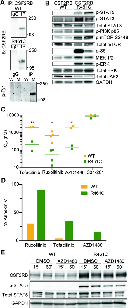
Figure 2. CSF2RB R461C is constitutively phosphorylated and activates canonical downstream pathways that are sensitive to JAK2 inhibition.
(A) CSF2RB was immunoprecipitated from transformed Ba/F3 lysates starved overnight in 0.1% FBS and probed for CSF2RB or phosphorylated tyrosine residues on replicate blots (W – WT lysate, M – R461C mutant lysate). WT cells were grown in IL-3 supplemented media. (B) Immunoblot analysis for activation of canonical CSF2RB signaling members across biologically replicate lysates. WT cells were grown in IL-3 supplemented media and all lines were starved overnight in 0.1% FBS. (C) Biologically replicate WT and R461C lines were screened on a small molecule inhibitor library and the top three hits (determined by % IC50 difference between the two lines) were the JAK-inhibitors Tofacitinib, Ruxolitinib and AZD1480. The STAT3 inhibitor S31-201 was equally ineffective on both WT and R461C (*p<0.05, **p<0.01). WT cells were grown and tested in IL-3 supplemented media. Key inhibitors are validated in Supplemental Figure 6A–C, full inhibitor panel results are found in Supplemental Table 3. (D) Lines were treated with indicated JAK-inhibitors (Ruxolitinib 350nM, Tofacitinib 350nM, AZD1480 1µM) for 48 hours and analyzed for apoptotic induction using Annexin V staining and flow cytometry (Guava PCA). 24 hour reads are shown in Supplemental Figure 6D. (E) Cells were starved overnight in 0.1% FBS and then treated with the JAK2 specific inhibitor AZD1480 (500nM) for the indicated time and immunoblotted for CSF2RB downstream signaling. WT cells were maintained in IL-3 supplemented media prior to starvation and treatment.

