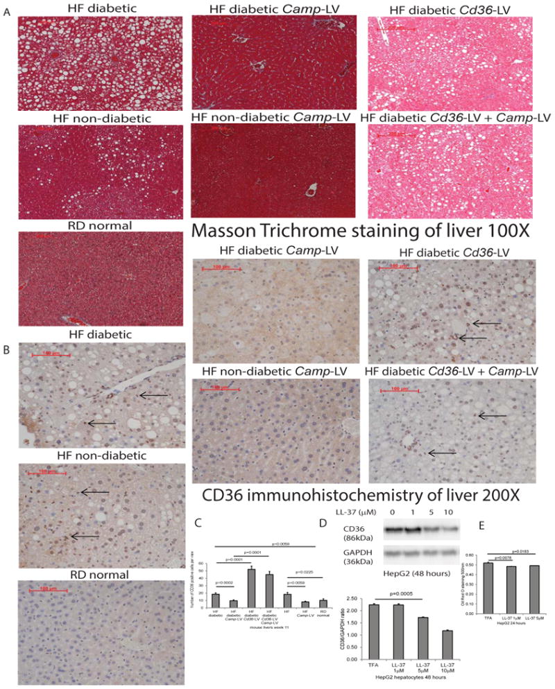
Figure 4. Lentiviral cathelicidin overexpression inhibited hepatic steatosis via CD36.

(A) Masson Trichrome staining of liver tissues of mice dissected on week 11. The background was stained in red color. Lipid accumulation was presented as white dots. High-fat diet-treated diabetic and non-diabetic mice all developed steatosis, compared with normal mice with regular diet. Steatosis was ameliorated in Camp-expressing groups that were reversed by Cd36 overexpression, regardless of diabetic status. (B) Immunohistochemistry of CD36 protein in liver tissues of mice dissected on week 11. (C) Quantification of CD36 protein in liver tissues. CD36 protein expression, as indicated by brown color, was significantly increased in high-fat diet-treated diabetic and non-diabetic mice, compared with normal mice with regular diet. Hepatic CD36 protein expression was significantly reduced in Camp-expressing groups, compared with control counterparts. CD36 protein expression was significantly increased in Cd36-expressing groups, indicating successful lentiviral transfection. n=6 mice per group. (D) Serum-starved HepG2 cells were exposed to TFA (vehicle control) or LL-37 (1-10 μM) for 24 hours. Western blot of HepG2 cells with quantification. LL-37 (1-10 μM) significantly reduced CD36 protein expression in human hepatocytes. (E) Oil red O staining was used to determine lipid accumulation. LL-37 (1-5 μM) significantly reduced lipid accumulation in hepatocytes. Experiments are representative of 2 independent experiments.
