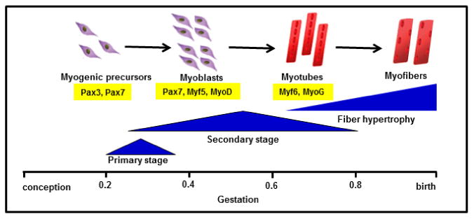
Figure 2. Schematic representation of fetal myogenesis during pregnancy.

The fraction of gestation when primary (embryonic) and secondary (fetal) stages of myogenesis are based on studies from sheep, cow, and human (Du et al., 2010, Romero et al., 2013, Wilson et al., 1992). Pax transcription factors Pax3 and Pax7 define the progenitor cell population during fetal myogenesis (Wang et al., 2010). Pax7 is expressed in fetal myoblasts, in addition to Myf5 and MyoD which commit cells to the myogenic program. Expression of the terminal differentiation genes, Myf6 (also known as Mrf4) and MyoG, are expressed in myotubes(Bentzinger et al., 2012). Myofibers grow by hypertrophy late in gestation and in postnatal life.
