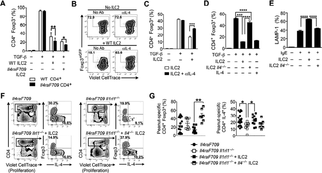
Figure 7. IL-4 secretion by ILC2 promotes food allergy by decreasing allergen-specific Treg cell induction.
(A) TGF-β induction of iTreg cells from naïve CD4+ WT or Il4raF709 T cells co-cultured with cell-sorted WT or Il4raF709 ILC2. (B) Flow cytometric analysis of the effect of ILC2 and anti(α)-IL-4 treatment on TGF-β-driven in vitro iTreg cell induction. (C) Frequency of TGF-β-induced iTreg cells co-cultured in the presence of WT ILC2 without or with αIL4. (D) Frequency of TGFβ-induced iTreg cells co-cultured with either WT ILC2, Il4−/− ILC2 or recombinant IL-4. (E) IgE-mediated mast cell degranulation following co-culture with ILC2, ILC2 supernatants or IL-4 in the presence or absence of αIL-4. (F) IgE-mediated degranulation of mast co-cultured with WT ILC2 or Il4−/− ILC2. (G) Flow cytometric analysis of peanut extract-induced in vitro proliferation of CD4+ Foxp3+ Treg cells and CD4+ Foxp3− T conventional cells isolated from the MLN of peanut-sensitized Il4raF709 and Il4raF709 Il1rl1−/− mice reconstituted with either WT or Il-4−/− ILC2 (F) Frequency of peanut-specific CD4+ Foxp3+ Treg cells and CD4+ T cells producing IL-4. Results represent data of 5 mice per group (A to F) and 7 to 18 mice (G and H) derived from two independent experiments. *P < .05, **P < .01, ***P < .001, ****P < .0001 by two-way and one-way ANOVA with post-tests analysis.

