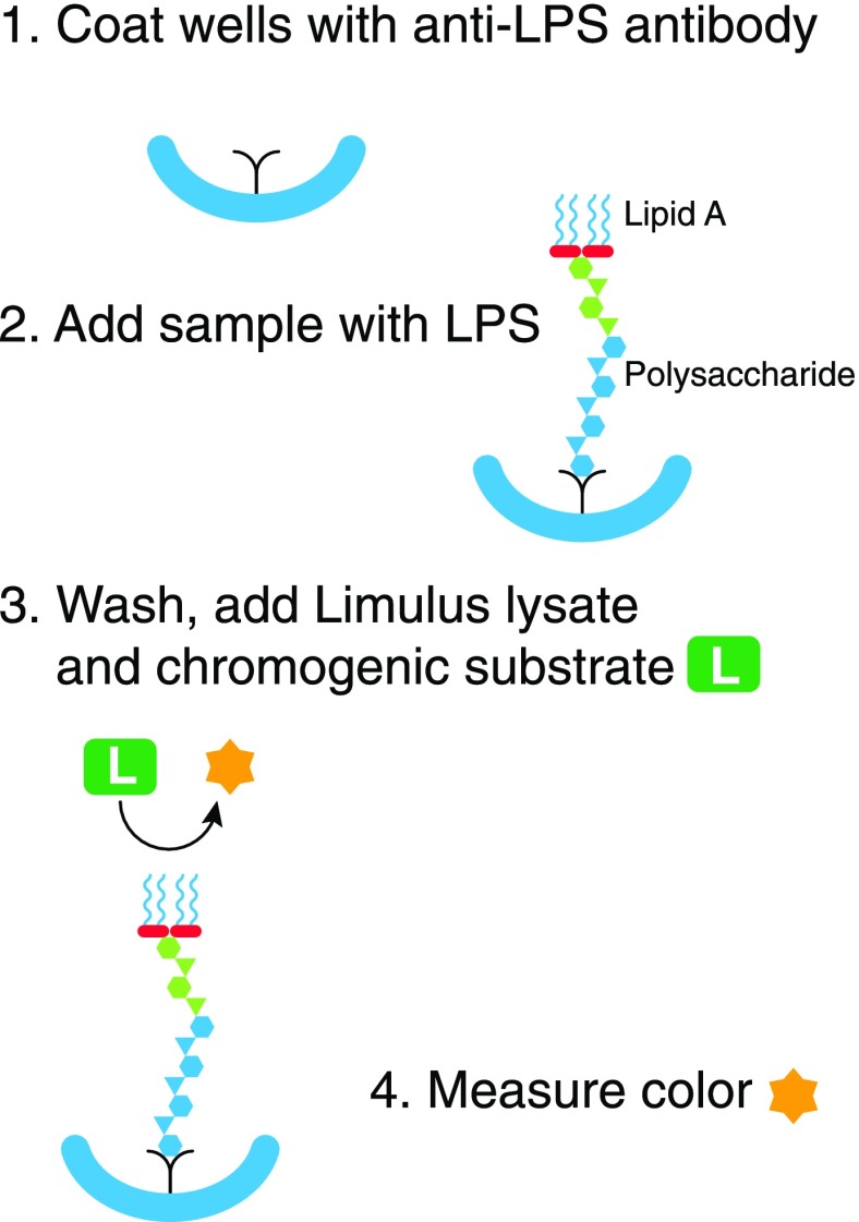
Figure 4. The “immunolimulus” assay.
Microtiter wells are first coated with an antibody to the target LPS (1). After washing, plasma is added. LPS in the plasma binds to the antibody (2). After another thorough wash, Limulus lysate and a chromogenic peptide substrate are added (3). The color produced is measured over time (4). The approach makes the LAL assay more specific for LPS.

