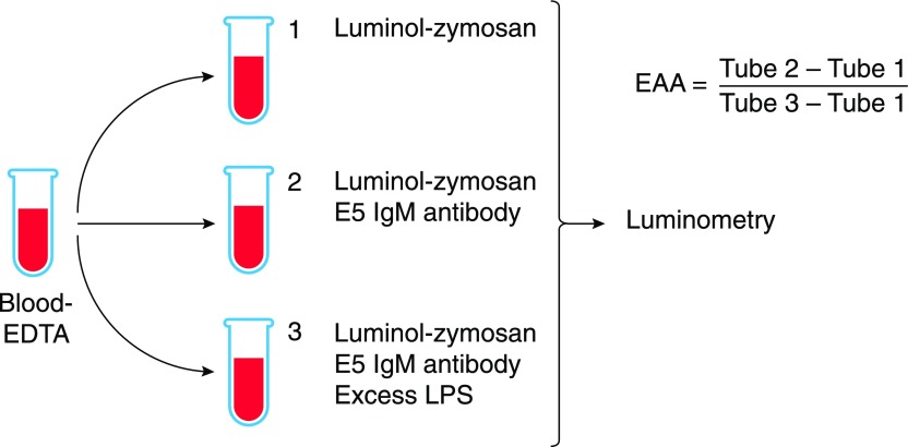
Figure 5. The EAA.
Anticoagulated blood is incubated with an excess of LPS in tube 3 before E5 IgM antibody is added to tubes 2 and 3 and luminol-zymosan is added to all tubes. After incubation at 37°C with shaking, photons are counted using a luminometer. EAA is calculated using the formula shown. Graphic adapted from Matsumoto et al. [85].

