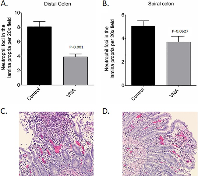
FIG 6.
Evaluation of neutrophilic foci and histopathologic lesions in the colon and large intestine. (A and B) Quantitative evaluation of neutrophilic foci in distal colon (A) and spiral colon (B) of untreated control and VNA-treated piglets. (C) Untreated piglets with mucosal ulceration, hemorrhage, and marked neutrophilic infiltration and eruption of neutrophils and sloughed mucosa into the intestinal lumen. (D) Treated piglets with mild mucosal erosion and neutrophilic infiltration.

