
Fig. 1. eNOS plays an essential role in retinal angiogenesis.
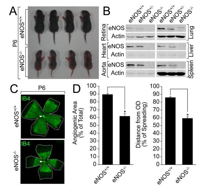
(A)A comparison of the body size between the wild type and eNOS knockout littermates was visualized by a digital camera at P6. (B) Lung, liver, spleen, heart, aorta, and retina were isolated from the wild type or eNOS-deficient mice, and the expression of eNOS was verified by a Western blot analysis with the indicated antibodies. (C and D) Retinas were isolated from the wild type and eNOS knockout mice at P6 and stained with IB4 (green). Angiogenesis was analyzed by measuring the angiogenic area and distance. Images were captured on confocal microscope at ×2.5 (zoom ×0.5) magnification. Angiogenic area and sprouting distance were quantified using Image J (National Institutes of Health, MD, USA) software. White arrowheads indicate optic nerve (ON). Data are presented as the means±SEM. Asterisks indicate statistical significance (p<0.05). Scale bars, 800 µm.