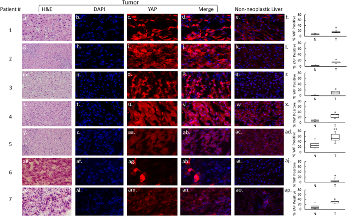
Figure 1. YAP nuclear localization is increased in moderately differentiated pediatric HCC compared to non-neoplastic liver.
Representative sections from tumor and matched non-neoplastic liver are shown at 40x (panels a–e, g–k, m–q, s–w, y–ac, ae–ai, ak–ao). The quantitative analysis of YAP nuclear staining for each sample is shown in panels f, l, r, x, ad, aj, and ap. YAP expression in the nuclei of tumor cells is increased for all patients. *p = 0.01; **p = 0.05.

