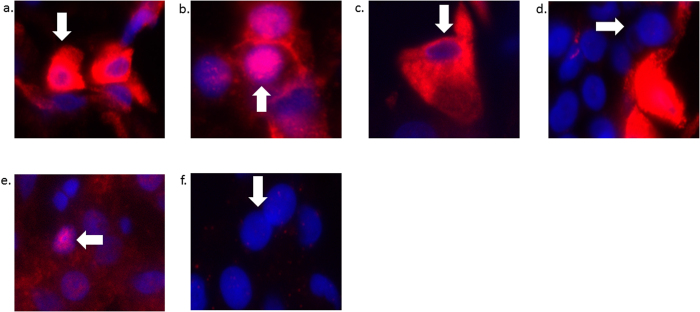
Figure 2.
Representative images of the different staining patterns for YAP: (a) combined nuclear and cytoplasmic YAP staining in tumor cells (white arrow), (b) nuclear-only staining in tumor cells, (c) cytoplasmic-only staining in tumor cells, (d) no staining in tumor cells, (e) nuclear staining in non-neoplastic cells, and (f) no staining in non-neoplastic cells. All images were taken at 40x, then cropped and expanded to display representative cells.

