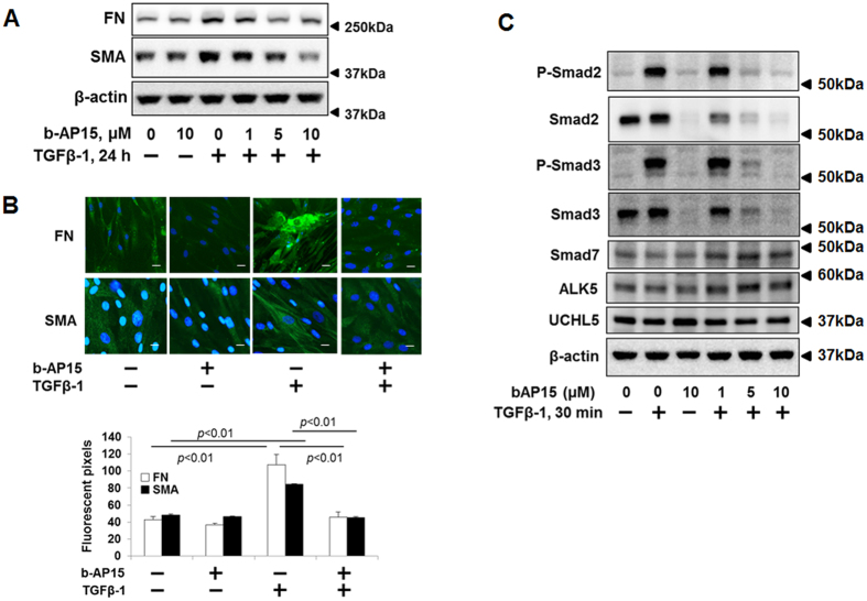
Figure 1. b-AP15 attenuates TGFβ-1 signaling in HLF cells.
(A) HLF cells were pre-treated with increasing doses of b-AP15 (0, 1, 5, and 10 μM) for 1 h, and then cells were treated with TGFβ-1 (2 ng/ml) for 24 h. Cell lysates were analyzed by immunoblotting with the antibodies to FN, α-SMA, and β-actin. (B) HLF cells were pre-treated with DMSO and b-AP15 (50 μM) for 1 h followed by TGFβ-1 (2 ng/ml) for 24 h, and then cells were fixed with 3.7% formaldehyde. Expressions of FN and α-SMA were detected by immunostaining with antibodies to α-SMA and FN. DAPI was used for nuclei staining (blue). Scale bar, 50 μm. (C) HLF cells were pre-treated with increasing doses of b-AP15 (0, 1, 5, and 10 μM) for 1 h followed by TGFβ-1 (2 ng/ml) for 30 minutes. Cell lysates analyzed by immunoblotting with the antibodies to p-Smad2, p-Smad3, Smad2, Smad3, Smad7, ALK5, UCHL5, and β-actin. Western blot images were cropped to improve the conciseness of the data; samples derived from the same experiment and the blots were processed in parallel. Representative of experiments performed at least 3 independent times.

