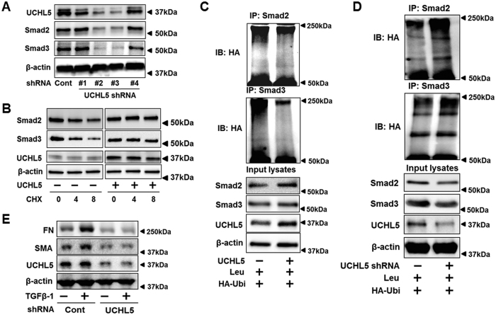
Figure 4. UCHL5 de-ubiquitinates and stabilizes Smad2/Smad3, and promotes TGFβ-1 signaling.
(A) HLF cells were co-transfected with scramble shRNA and four different designed UCHL5 shRNA (#1–#4) for 3 days, and then cell lysates were analyzed by immunoblotting with the antibodies to UCHL5, Smad2, Smad3, and β-actin. (B) MLE12 cells transfected with empty vector plasmid or UCHL5 plasmid for 48 h, and cells were treated with CHX (20 μg/ml) for 0–8 h. Cell lysates were analyzed by immunoblotting with the antibodies to Smad2, Smad3, UCHL5, and β-actin. (C) HLF cells were co-transfected with HA-Ubi or HA-Ubi+UCHL5 plasmids for 48 h. Cell lysates were subjected to immunoprecipitation with Smad2 or Smad3 antibody, followed by immunoblotting with HA tag antibody. Input lysates were analyzed by immunoblotting with Smad2, Smad3, UCHL5, and β-actin. (D) HLF cells were transfected with scramble (cont) shRNA or UCHL5 shRNA, and then transfected with HA-Ubi plasmids for 48 h. Cell lysates were subjected to immunoprecipitation with Smad2 or Smad3 antibody, followed by immunoblotting with HA tag antibodies. Input lysates were analyzed by immunoblotting with Smad2, Smad3, UCHL5, and β-actin. (E) HLF cells were transfected with cont shRNA and UCHL5 shRNA for 72 h, and then cells were treated with TGFβ-1 (2 ng/ml) for 24 h. Cell lysates were analyzed by immunoblotting with the antibodies to FN, α-SMA, UCHL5, and β-actin. Western blot images were cropped to improve the conciseness of the data; samples derived from the same experiment and the blots were processed in parallel. Representative of experiments performed at least 3 independent times.

