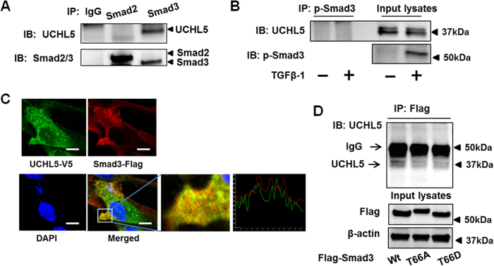
Figure 5. UCHL5 is associated with Smad3.
(A) HLF cell lysates were subjected to immunoprecipitation with antibodies to IgG, Smad2, or Smad3, followed by immunoblotting with UCHL5 and Smad2/Smad3 antibodies. (B) HLF cells were treated with TGFβ-1 (2 ng/ml) for 30 min. Cell lysates were subjected to immunoprecipitation with an antibody to p-Smad3, followed by immunoblotting with UCHL5 antibody. Input lysates were analyzed by immunoblotting with p-Smad3 antibody. (C) HLF cells were co-transfected with Flag-Smad3 and V5-tagged UCHL5 (UCHL5-V5) for 48 h, and then cells were fixed with 3.7% formaldehyde. Localization of Flag-Smad3 and UCHL5-V5 were detected by co-immunostaining with antibodies to Flag tag (red) and V5 tag (green). Nuclei were stained with DAPI (blue). Scale bar, 10 μm. 92.0% of the cells with double staining show positive co-localization. (D) HLF cells were transfected with Flag-Smad3 wild type (Wt), Flag-Smad3T66A, and Flag-Smad3T66D plasmids for 48 h, and then cell lysates were subjected to immunoprecipitation with an antibody to Flag tag, followed by UCHL5 immunoblotting. Input lysates were analyzed by immunoblotting with Flag tag and β-actin antibodies. Western blot images were cropped to improve the conciseness of the data; samples derived from the same experiment and the blots were processed in parallel. Representative of experiments performed at least 3 independent times.

