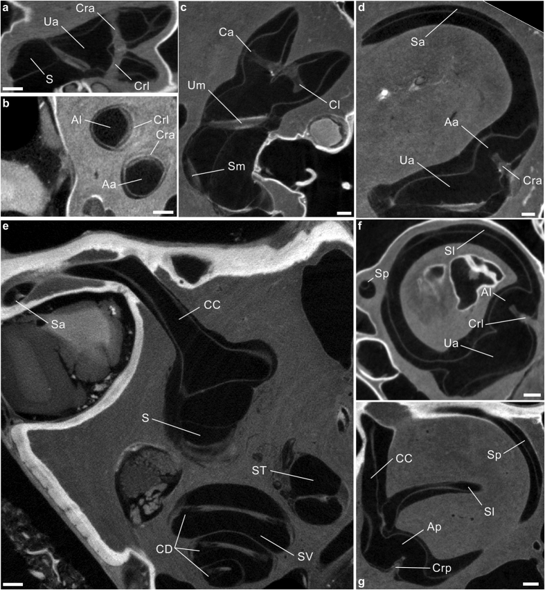
Figure 1. Raw visualizations of stained membranous labyrinths through micro-CT scanning.
(a–g) Soft-tissues structures contained inside the bony labyrinth of the human (c,d), rhesus macaque (b,e,g) and squirrel monkey (a,f) specimens. Aa, Ap, Al, anterior, posterior and lateral ampullae, respectively; Ca, Cl, anterior and lateral cupulae (artificially shrunk); CC, common crus; CD, cochlear duct; Cra, Crp, Crl, anterior, posterior and lateral cristae ampullares; S, sacculus; Sa, Sp, Sl, slender parts of the anterior, posterior and lateral semicircular ducts; Sm, macula sacculi; ST, scala tympani; SV, scala vestibuli; Ua, anterior utriculus; Um, macula utriculi. Scale bar is 0.5 mm.

