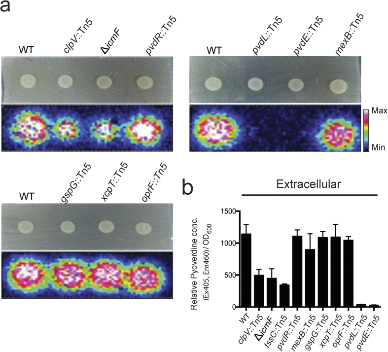
Figure 6. MALDI-IMS analysis and quantification of extracellular mature pyoverdine.
(a) MALDI-IMS analysis of secretory mature pyoverdine (m/z 1044) on the surface of iron-limited agar plates of wild-type, T6SS clpV::Tn5, T6SS ΔicmF, T2SS gspG::Tn5, T2SS xcpT::Tn5, PvdRT-OmpQ pvdR::Tn5, MexAB-OprM mexB::Tn5, outer membrane protein ΔoprF::Tn5 mutants. Bacteria were grown for 12 h on the iron-limited agar plates. (b) Quantification of extracellular mature pyoverdine was detected by measuring fluorescence at excitation 405 nm and emission 460 nm. Pyoverdine values were normalized against the cell optical densities (Ex 405, Em 460/OD600), and relative values were determined by comparing each value to the periplasmic mean value of the wild-type. Values are mean ± SD of three independent experiments. Bacteria were grown on the iron-limited media for 12 h at 28 °C.

