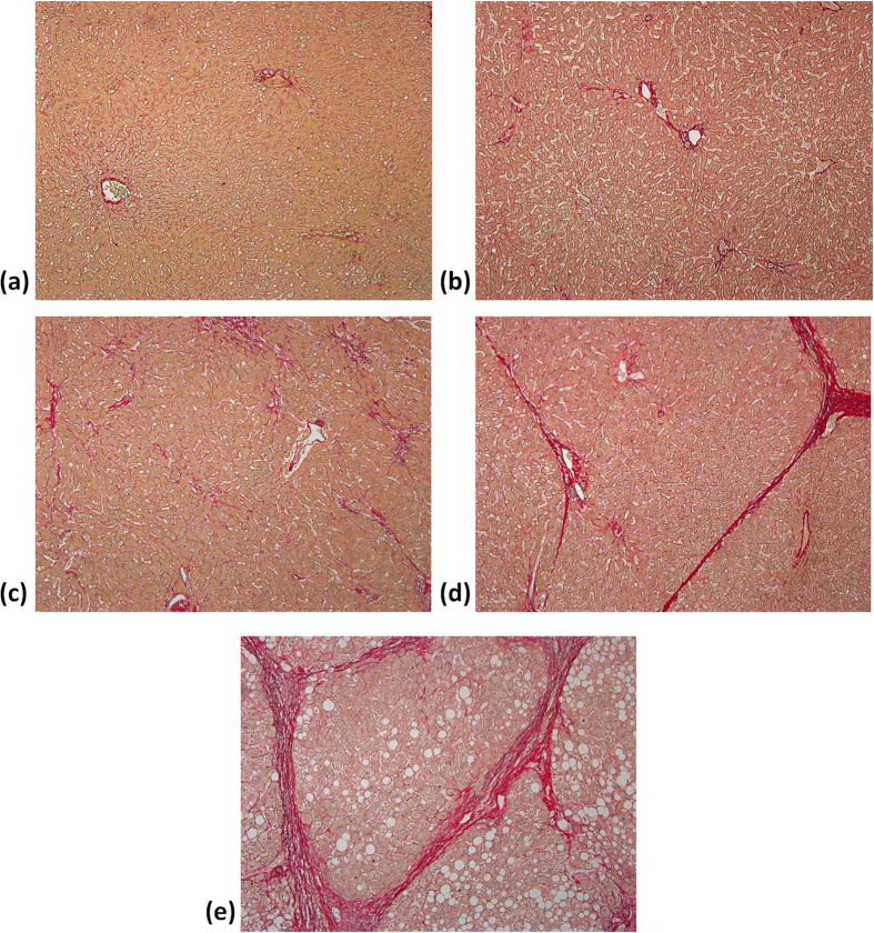
Figure 1. PR-stained sections (100 magnification) obtained from patients’ livers with different degrees of fibrosis from F0 to F4.
(a) F0: no fibrosis; (b) F1: fibrous expansion of portal areas without septa (i.e., portal fibrosis); (c) F2: portal fibrosis with few septa was observed; (d) F3: fibrous expansion of portal areas with marked bridging or septa (i.e., septal fibrosis); (e) F4: the tissue is composed of nodules surrounded completely by fibrosis (i.e., cirrhosis).

