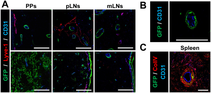
Figure 5. ColVI-Cre mice target perivascular cells in SLOs.
(A) Representative images of T- (top) and B- (bottom) cell areas in PPs, pLNs and mLNs, showing Cre-mediated GFP expression in relation to blood and lymphatic vessels. BECs are marked as CD31+ cells (blue) and LECs as CD31+ Lyve-1+ cells (purple). (B) Higher magnification image of a blood vessel (CD31+) and GFP+ surrounding it. (C) Spleen image showing GFP+ cells only in the central part of the white pulp, surrounding endothelial cells (CD31+ cells in blue) and co-localizing with collagen IV (red). Scale bar, 75 μm. 5 different mice were analyzed.

