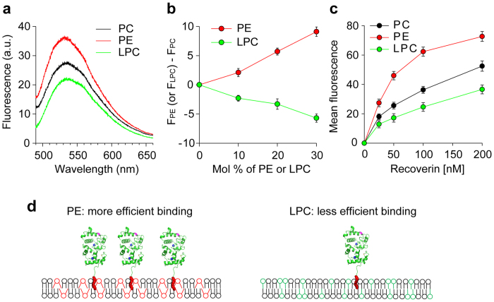
Figure 3. Effect of spontaneous membrane curvature on membrane association of recoverin.
(a) Fluorescence emission spectra of 0.1 μM mRec-F23NBD were recorded in the presence of LUVs (0.1 mM total lipids) composed of PC, PC:PE (7:3), or PC:LPC (7:3) and in the presence of 1 mM Ca2+. (b) Relative fluorescence intensities measured as a function of the mol fractions of PE (red) or LPC (green) in PC bilayers at excitation and emission wavelengths of 475 and 530 nm, respectively. (c) Mean fluorescence intensity recorded by TIRF microscopy for the binding of mRec-F158NBD to supported membranes composed of PC, PC:PE (7:3), or PC:LPC (7:3) in the presence of 1 mM Ca2+. (d) Schematic diagram of lipid-dependent association of recoverin to membranes. Recoverin binds more efficiently to membranes containing cone-shaped lipids (PE) than inverted cone-shaped lipids (LPC). Data are representative of three experiments in a, and mean ± s.d. of triplicates in b and c.

