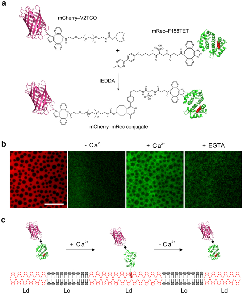
Figure 5. Synthesis of mCherry-recoverin conjugate and its Ca2+-responsive translocation to phase-separated supported membrane.
(a) Schematic diagram of coupling between mCherry-V2TCO and mRec-F158TET. (b) Ca2+-dependent binding of the mCherry-recoverin conjugate to a supported membrane with ordered and disordered lipid domains. Supported membranes (left) were composed of DPPC:DOPC:Cholesterol (2:2:1) with coexisting Lo (dark) and Ld (red) phases. The membranes were labeled with 0.5 mol% DiD, which preferentially partitions into the Ld phase. The mCherry-recoverin conjugate was added to the membranes in the absence (center left) or in the presence (center right) of Ca2+. Addition of 2 mM EGTA (right) extracts most of the membrane-bound conjugate. Scale bar is 20 μm. Representative images of three experiments. (c) Schematic diagram depicting the reversible translocation of mCherry via the Ca2+-sensitive myristoyl chain (red) of recoverin into fluid phase regions of a phase-separated Lo/Ld membrane.

