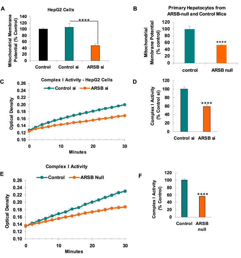
Figure 4. Mitochondrial membrane potential and Complex 1 activity in ARSB-null mice.
(A) Mitochondrial membrane potential was measured in the HepG2 cells and shown to be significantly less when ARSB was silenced (n = 6; one-way ANOVA with Tukey-Kramer post-test). (B) Similarly, the mitochondrial membrane potential in the mitochondria from the ARSB-null primary hepatocytes was significantly less than in hepatocytes from the C57BL/6J control mice (n = 6). (C) The activity of Complex 1 was determined by an NADH dehydrogenase activity assay which showed marked reduction of activity in the mitochondria of the HepG2 cells following ARSB silencing, compared to control (n = 6). (D) Graphical representation of the slope of the activity shows the significant difference following ARSB silencing. (E) Similarly, Complex 1 activity was markedly reduced in the crude mitochondrial fraction from ARSB-null mouse hepatic tissue, compared to the value in the C57BL/6J (n = 6). (F) Graphical representation of the decline in Complex 1 activity of the ARSB-null mouse hepatic tissue, compared to the control [ARSB = arylsulfatase B; ER = endoplasmic reticulum; MMP = mitochondrial membrane potential].

