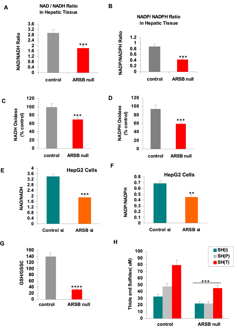
Figure 6. NAD+/NADH and NADP+/NADPH ratios, NADH and NADPH oxidase activity, and GSH/GSSG ratio and thiol content.
(A) The ratio of NAD+ to NADH is significantly lower in the ARSB-null mouse hepatic tissue, mainly due to increase in NADH, which increased by ~66 ng/mg protein in the ARSB-null mouse hepatic tissue (n = 20). (B) In the ARSB-null mouse hepatic tissue, the ratio of NADP+ to NADPH was also less, largely attributable to an increase in NADPH of ~9.5 ng/mg protein (n = 20). (C) Consistent with the increase in NADH, the NADH oxidase was significantly less in the ARSB-null mice (n = 12). (D) Consistent with the observed increase in NADPH, the NADPH oxidase activity was less in the ARSB-null hepatic tissue (n = 20). (E) In the ARSB-silenced HepG2 cells, the ratio of NAD+ to NADH was also significantly less, due predominantly to increase of ~80 ng/mg protein in NADH (n = 3). (F) In the ARSB-silenced HepG2 cells, the ratio of NADP+ to NADPH was significantly less, due to increase of ~8 ng/mg protein in NADPH (n = 3). (G) In contrast to the increases in the reduced forms NADH and NADPH when ARSB was lower in the ARSB-null mouse hepatic tissue, the ratio of glutathione (GSSG) to reduced glutathione (GSH) was increased, due to decline in reduced glutathione (n = 12). (H) Similarly, the level of thiols was significantly reduced in the ARSB-null hepatic tissue, demonstrating an overall decline in reduced sulfur products when ARSB activity was less (n = 12). [ARSB = arylsulfatase B; CF = control female; CM = control male; GSH = glutathione; GSSG = glutathione disulfide].

