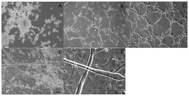
Figure 1.
Phase images of myoblasts and myotubes derived from the Tibalias anterior of adult Sprague Dawley rats. Phase images of proliferating myoblasts at (A) 2 DIV, (B) 4 DIV, (C) 8 DIV. Phase images of fused myoblasts at 12 DIV (D,E). Scale Bar 50 μm, the scale bar in panel D pertains to panels A–D.

